PHLEBOTOMY (BLOOD DRAW)

Premier’s On-site Phlebotomy Patient Service Centers allow our patients a convenient way to get the tests that they need as quickly as possible. It also means your results are sent directly to your physician without any delays.

No appointment is necessary and walk-ins are welcome;  however, tests must be ordered by your Premier Medical Group provider.

To better serve our patients,  in-house phlebotomy services at the following locations:

Premier Medical Clinic

Monday – Friday: 8:00 am – 4:00 pm
(The lab is closed 12:00 pm – 1:00 pm each day for lunch)

+850-584-3278

Please do not contact the patient lab directly for test results.

Please allow 2 weeks from when your blood work is drawn, for results to be available.

If you do not receive some type of communication from the ordering provider regarding your results please contact their office.

Premier Medical Group

PATHOLOGY & CLINICAL LABS

Premier Medical Group is proud to have in-house clinical and pathology labs.  Having an in-house lab guarantees that Premier’s labwork is done promptly and efficiently so our patients won’t have to endure anxious waits to learn about the next step in their treatment.

The close relationship with the in-house labs gives our physicians the opportunity to communicate directly with the lab’s tech staff. We are also able to work with our pathologist to customize testing parameters that provide information that may be more meaningful to the physician, and patient treatment, than the standard protocol.

Premier Medical Group

RADIOLOGY

RADIOLOGY AT PREMIER MEDICAL GROUP

For the convenience of our patients, Premier Medical Group offers many diagnostic imaging services in-house. Quality, service and patient satisfaction is Premier Medical Group’s top priority.

RADIOLOGY SERVICES OFFERED:

X-Ray

An X-ray is a quick, painless test that produces images of the structures inside your body — particularly your bones.

X-ray beams pass through your body, and they are absorbed in different amounts depending on the density ofthe material they pass through. Dense materials, such as bone and metal, show up as white on X-rays. The air in your lungs shows up as black. Fat and muscle appear as shades of gray.

For some types of X-ray tests, a contrast medium — such as iodine or barium — is introduced into your body to provide greater detail on the images.

What you can expect:   An x-ray technologist will position your body to best achieve the desired view. While the x-ray is being taken, you will remain perfectly still so that the resulting picture is clear. An X-ray procedure may take just a few minutes for a simple X-ray or longer for more-involved procedures, such as those using a contrast medium.

Computerized Tomography (CT Scan)

A computerized tomography (CT) scan combines a series of X-ray images taken from different angles around your body and uses computer processing to create cross-sectional images (slices) of the bones, blood vessels and soft tissues inside your body. CT scan images provide more-detailed information than plain X-rays do.

A CT scan can be used to visualize nearly all parts of the body and is used to diagnose disease or injury as well as to plan medical, surgical or radiation treatment.

What you can expect: CT scanners are shaped like a large doughnut standing on its side. You lie on a narrow, motorized table that slides through the opening into a tunnel. Straps and pillows may be used to help you stay in position. During a head scan, the table may be fitted with a special cradle that holds your head still.

While the table moves you into the scanner, detectors and the X-ray tube rotate around you. Each rotation yields several images of thin slices of your body. You may hear buzzing and whirring noises.

A technologist in a separate room can see and hear you. You will be able to communicate with the technologist via intercom. The technologist may ask you to hold your breath at certain points to avoid blurring the images. The whole process typically takes about 30 minutes.

Ultrasound

Diagnostic ultrasound, also called sonography or diagnostic medical sonography, is an imaging method that uses high-frequency sound waves to produce images of structures within your body. The images can provide valuable information for diagnosing and treating a variety of diseases and conditions.

Most ultrasound examinations are done using an ultrasound device outside your body, though some involve placing a device inside your body.

What you can expect: During an ultrasound exam, gel is applied to the skin. A sonographer will press a small, hand-held device (transducer) against the area being studied and moves it as needed to capture the images. The transducer sends sound waves into your body, collects the ones that bounce back and sends them to a computer, which creates the images.

A typical ultrasound exam takes from 30 minutes to an hour.

MRI

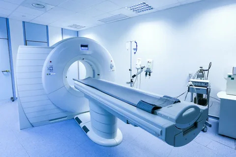
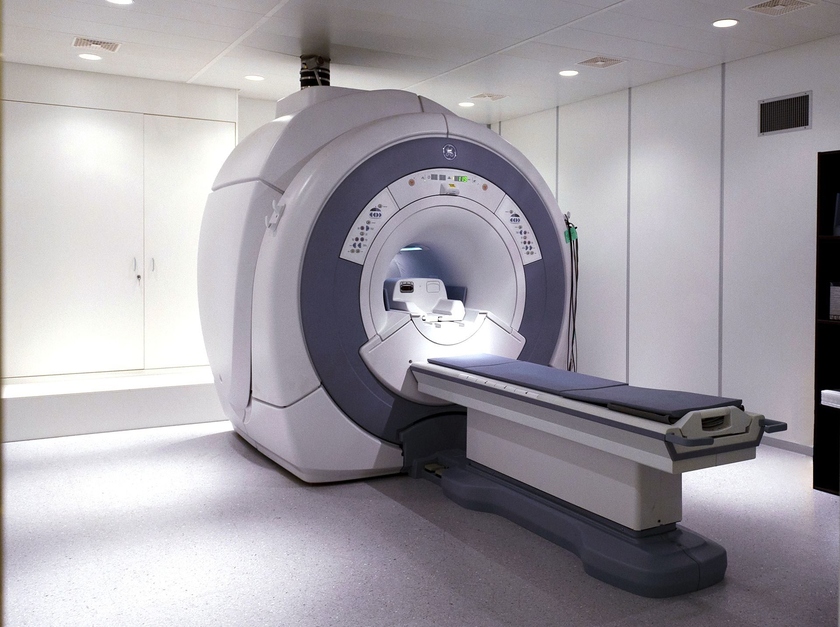
An MRI (magnetic resonance imaging) is a sophisticated diagnostic technique that is non-invasive and does not use radiation. The MRI uses a magnetic field, radio waves, and a computer to generate detailed 2-dimensional images rather than flat x-ray images. The MRI can be used to detect cancer in a variety of organs and tissues. The images are so clear that many organs can be seen in great detail. The MRI can also detect injuries, disorders, and diseases affecting tendons, ligaments, cartilage, and bone marrow.